Anatomy Of Musckes Sndctendons - Human Muscle System Functions Diagram Facts Britannica - Smooth muscles (involuntary muscles) are usually in sheets or layers, with one layer of muscle behind the other.
Anatomy Of Musckes Sndctendons - Human Muscle System Functions Diagram Facts Britannica - Smooth muscles (involuntary muscles) are usually in sheets or layers, with one layer of muscle behind the other.. There are two main muscle groups around the knee: Smooth muscles (involuntary muscles) are usually in sheets or layers, with one layer of muscle behind the other. There are around 650 skeletal muscles within the typical human body. Related online courses on physioplus. The three scalene muscles are found forming the floor of the posterior triangle.
The inguinal aponeurotic falx (falx aponeurotica inguinalis; Topographically, the muscles in this group are classed along with the lateral torso wall and upper extremity, which is due to their location as well as their genetic development based on their embryological origin. More serious injuries may result in partial or complete tear of the calf. Conjoined tendon of internal oblique and transversalis muscle) of the obliquus internus and transversus is mainly. See the pictures and anatomy description of knee joint bones, cartilage, ligaments, muscle and tendons with resources for knee problems & injuries.
Anatomy Of Knee from ix-cdn.b2e5.com Learn about human anatomy muscles with free interactive flashcards. Find the best weight lifting exercises that target each muscle or groups of muscles. Learning to draw muscles may conjure medical charts in daunting details, but such complexity is unnecessary. This fascicular organization is common in muscles of the limbs; Almost every muscle constitutes one part of a pair of identical bilateral. The anterior and middle scalenes originate from the transverse processes of certain cervical vertebrae and attach to the first rib. Discover the muscle anatomy of every muscle group in the human body. See the pictures and anatomy description of knee joint bones, cartilage, ligaments, muscle and tendons with resources for knee problems & injuries.
As the skeletal muscles pull on bones to cause movements, they also stabilize the joints of the skeleton;
The inguinal aponeurotic falx (falx aponeurotica inguinalis; See more ideas about muscle anatomy, anatomy, hip muscles anatomy. The anterior and middle scalenes originate from the transverse processes of certain cervical vertebrae and attach to the first rib. The tendons of these muscles pass through a small corridor in the wrist known as the carpal tunnel. There are around 650 skeletal muscles within the typical human body. In this section, learn more about the anatomy of the muscles of the neck. The muscle groups of the upper leg region are the gluteal group. The anatomy of muscle cells differs from that of other body cells and biologists have applied specific terminology to different parts of these cells. Understanding the structure of a muscle fiber. The muscles of the abdomen may be divided into two groups: Microscopic anatomy of skeletal muscle. More serious injuries may result in partial or complete tear of the calf. A calf muscle strain is commonly called a pulled calf muscle.
The anatomy of muscle cells differs from that of other body cells and biologists have applied specific terminology to different parts of these cells. • the muscular system develops from intra embryonic mesoderm. The muscle groups of the upper leg region are the gluteal group. This article will focus on tongue embryology, origin, insertion, and action of the extrinsic muscles, followed by innervation, blood supply and lymphatic drainage of the tongue. • definitions • introduction • development of muscles • classification • anatomy of skeletal muscle • muscle physiology • properties • muscles of development of muscles.
Dog Anatomy Muscles And Tendons The Y Guide from www.caninefitness.com There are around 650 skeletal muscles within the typical human body. The three scalene muscles are found forming the floor of the posterior triangle. It allows the nervous system to trigger a specific movement of a muscle by. You can click the links in the image, or the links below the image to find out more information on any muscle group. Topographically, the muscles in this group are classed along with the lateral torso wall and upper extremity, which is due to their location as well as their genetic development based on their embryological origin. The muscles of the abdomen may be divided into two groups: Almost every muscle constitutes one part of a pair of identical bilateral. Learn about the muscles, tendons, bones, and ligaments that comprise the knee joint anatomy.
See the pictures and anatomy description of knee joint bones, cartilage, ligaments, muscle and tendons with resources for knee problems & injuries.
Knee function is determined in large part by the anatomy of the joint. More serious injuries may result in partial or complete tear of the calf. Topographically, the muscles in this group are classed along with the lateral torso wall and upper extremity, which is due to their location as well as their genetic development based on their embryological origin. Anatomy of the muscular system. • muscle tissues develop from embryonic cells. All calf muscle strains result in tearing of some muscle fibers. Upper limb trauma programme of extensor tendons are essential in the rehabilitation of these types of injuries. See the pictures and anatomy description of knee joint bones, cartilage, ligaments, muscle and tendons with resources for knee problems & injuries. Muscular contraction is necessary for voluntary and involuntary movement of limbs, stabilization of joints, maintaining luminal diameter (in the case of arteries, bowel, etc), and to produce heat. Convergent muscles contain fibers that have a wide origin, but converge in order to attach to a narrow tendon. Find the best weight lifting exercises that target each muscle or groups of muscles. Related online courses on physioplus. This fascicular organization is common in muscles of the limbs;
Skeletal muscles allow the body to move and maintain posture; See the pictures and anatomy description of knee joint bones, cartilage, ligaments, muscle and tendons with resources for knee problems & injuries. The muscles around the knee help to keep the knee stable, well aligned, and moving. In the diagrams below, i'll be showing muscle groups in color, with a black line to show the forms that would show through the skin (i also show protruding bones that would do the same). The muscles of the abdomen may be divided into two groups:
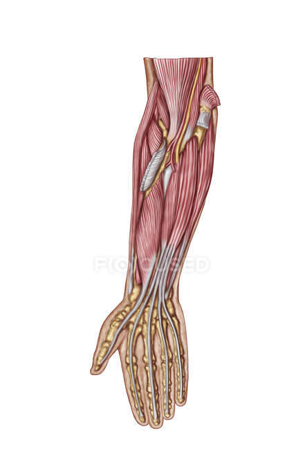
Anatomy Of Human Forearm Muscles Tendons Flexor Carpi Ulnaris Stock Photo 174718252 from st.focusedcollection.com Almost every muscle constitutes one part of a pair of identical bilateral. There's no strict demarcation or dividing line between the tendon and the covering around this muscle but that covering is called is called the epimysium fp my cm and it's really just connective tissue that covers the muscle kind of protects it reduces friction. Smooth muscles (involuntary muscles) are usually in sheets or layers, with one layer of muscle behind the other. Inside each skeletal muscle, muscle fibers are organized into bundles, called fascicles, surrounded by a middle layer of connective tissue called the perimysium. This fascicular organization is common in muscles of the limbs; • definitions • introduction • development of muscles • classification • anatomy of skeletal muscle • muscle physiology • properties • muscles of development of muscles. Muscle mass accounts for a large majority of the carcass weight of domestic animals. See the pictures and anatomy description of knee joint bones, cartilage, ligaments, muscle and tendons with resources for knee problems & injuries.
Conjoined tendon of internal oblique and transversalis muscle) of the obliquus internus and transversus is mainly.
Attached to the bones of the skeletal system are about 700 named muscles that make up roughly half of a person's body weight. Muscles of the thorax & abdomen | anatomy model. Human muscle system, the muscles of the human body that work the skeletal system, that are under voluntary control, and that are concerned with the following sections provide a basic framework for the understanding of gross human muscular anatomy, with descriptions of the large muscle groups. The muscular system consists of the skeletal muscles and their associated structures. Muscular contraction is necessary for voluntary and involuntary movement of limbs, stabilization of joints, maintaining luminal diameter (in the case of arteries, bowel, etc), and to produce heat. There are around 650 skeletal muscles within the typical human body. The three scalene muscles are found forming the floor of the posterior triangle. You can click the links in the image, or the links below the image to find out more information on any muscle group. By contracting, they also aid the venous return of blood to the heart and with age, these components of the musculoskeletal system progressively degenerate, which contributes to frailty and increases the risk of falls and fractures. Anatomy of the short head of the biceps brachii muscle. Muscles of the upper and lower leg. Convergent muscles contain fibers that have a wide origin, but converge in order to attach to a narrow tendon. • the muscular system develops from intra embryonic mesoderm.


0 Komentar